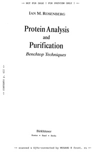
Protein Analysis and Purification
| Автор(ы): | Rosenberg J. M.
06.10.2007
|
| Год изд.: | 1996 |
| Описание: | This book is designed to be a practical progression of experimental techniques an investigator may follow when embarking on a biochemical project. The protocols may be performed in the order laid out or may be used independently. The aim of the book is to assist a wide range of researchers, from the novice to the frustrated veteran, in the choice and design of experiments that are to be performed to provide answers to specific questions. The manual describes standard techniques that have been shown to work, as well as some newer ones that are beginning to prove important. By following the prominently numbered steps, you can work your way through any protocol, whether it's a new technique or a task you've done before for which you need a quick review or updated methodology. |
| Оглавление: |
 Обложка книги.
Introduction [1] How to Use this Book [2] Basic Laboratory Equipment [3] Beyond Protein Analysis and Purification [4] Chapter 2 Protein Structure [5] Introduction [5] A.The Amino Acids [5] B. The Four Levels of Protein Structure [6] Primary Structure [6] Secondary Structure [10] Tertiary Structure [12] Quaternary Structure [13] C. Chemical Characteristics of Proteins [14] Hydrophobicity [15] Consensus Sequences [16] Chapters 3 Tracking the Target Protein [19] Introduction [19] A. Labeling Cells and Proteins [21] Metabolic Labeling Cells in Culture [22] PROTOCOL 3.1 Metabolic Labeling of Adherent Cells [23] PROTOCOL 3.2 Metabolic Labeling Cells Growing in Suspension [23] PROTOCOL 3.3 Pulse-Chase [24] Surface Labeling [25] PROTOCOL 3.4 Lactoperoxidase Labeling Cell Surface Proteins [26] PROTOCOL 3.5 Labeling Surface Proteins by the IODO-GEN Method [27] PROTOCOL 3.6 Surface Labeling with Biotin [28] PROTOCOL 3.7 Domain-Selective Biotinylation and Streptavidin-Agarose Precipitation [29] PROTOCOL 3.8 Labeling Isolated Proteins by the Chloramine Т Reaction [31] B. Lysis—Preparation of the Cell Free Extract [32] PROTOCOL 3.9 Lysis of Cells in Suspension (continuation of Protocol 3.2) [33] PROTOCOL 3.10 Lysis of Adherent Cells (continuation of Protocol 3.1) [33] C. Principles of Immunoprecipitation [34] Antibodies as Detection Tools [34] Polyclonal Antibodies [34] Monoclonal Antibodies [35] Antibody Based Analytical Techniques: Western Blotting and Immunoprecipitation [35] PROTOCOL 3.11 Immunoprecipitation [37] PROTOCOL 3.12 Sequential Immunoprecipitation—Dissociation and Reimmunoprecipitation of Immune Complexes [39] PROTOCOL 3.13 Nondenaturing Immunoprecipitation [40] D. Additional Methods to Identify Associated Proteins [41] Sucrose Gradients [41] PROTOCOL 3.14 Preparation of Sucrose Gradients [42] Fractionating a Sucrose Gradient [43] Chemical Cross-linking [46] PROTOCOL 3.15 Cross-linking Proteins Added to Cells: Analysis of Receptor-Ligand Interaction [49] PROTOCOL 3.16 Cross-linking Proteins in Solution [50] PROTOCOL 3.17 Cross-linking Extraneously Added Ligand to Cells [50] PROTOCOL 3.18 Cross-linking Proteins in Solution Using the Homobifunctional Reagent Dithiobis(succinimidyl propionate) (DSP) [51] PROTOCOL 3.19 Analysis of the Cross-linked Products: Elution of Proteins from the Gel [52] Chapter 4 Electrophoretic Techniques [55] Introduction to Polyacrylamide Gel Electrophoresis (PAGE) [55] A. Preparation of SDS-Polyacrylamide Gels [57] PROTOCOL 4.1 Assembling the Plates [57] Choosing the Acrylamide Concentration [59] PROTOCOL 4.2 Casting the Separating Gel [59] PROTOCOL 4.3 Casting the Stacking Gel [61] PROTOCOL 4.4 Preparing the Sample [61] PROTOCOL 4.5 Attaching the Gel Cassette to the Apparatus, Loading the Samples, and Running the Gel [63] PROTOCOL 4.6 Gradient Gels [66] PROTOCOL 4.7 Tube Gels [67] PROTOCOL 4.8 Drying the Gel [68] PROTOCOL 4.9 Separation of Low Molecular Weight Proteins by Tricine-SDS-PAGE (TSDS-PAGE) [69] Safety Considerations [77] B. Detection of Protein Bands in Polyacrylamide Gels [71] PROTOCOL 4.10 Staining and Destaining the Gel with Coomassie Blue [72] PROTOCOL 4.11 Silver Staining [73] PROTOCOL 4.12 Reversible Staining of Proteins in Gels with a Heavy Metal Salts (CuCl(?) and ZnCl(?)) [74] PROTOCOL 4.13 Molecular Weight Determination by SDS-PAGE [75] C. Recovery of Proteins from the Gel [77] PROTOCOL 4.14 Excising the Protein Band from the Dried Gel [77] PROTOCOL 4.15 Extracting the Protein from the Dried Gel [78] PROTOCOL 4.16 Trichloroacetic Acid (TCA ) Precipitation [79] PROTOCOL 4.17 Electroelution of Protein Bands from SDS-PAGE Using ELUTRAP™ [79] D. 2-Dimensional Gel Systems [81] PROTOCOL 4.18 Two-Dimensional Gels: Nonreducing and Reducing [81] Isoelectric Focusing [84] PROTOCOL 4.19 Preparation of the Sample for Isoelectric Focusing [84] PROTOCOL 4.20 Preparation of the Isoelectric Focusing Gel Mixture [86] PROTOCOL 4.21 Measuring the pH of the Gel Slices [88] PROTOCOL 4.22 Nonequilibrium pH Gradient Electrophoresis (NEPHGE) [88] PROTOCOL 4.23 2-D Gels—The Second Dimension: SDS-PAGE [90] E. Identification of Enzyme Activity in Polyacrylamide Gels [90] General Considerations [91] PROTOCOL 4.24 Localization of Proteases—Copolymerization of Substrate in the Separating Gel [91] PROTOCOL 4.25 Identification of Protease Inhibitors—Reverse Zymography [92] PROTOCOL 4.26 Reacting the Gel with Solution after Electrophoresis [93] Identification of DNA Binding Proteins—Gel Shift Assay [94] PROTOCOL 4.27 Gel Shifts [95] Chapter 5 Getting Started with Protein Purification [99] Introduction [99] A. Making a Cell Free Extract [101] Cellular Disruption [102] Extraction Buffer Composition [102] Protease Inhibitors [102] Methods of Cell Disruption [103] Clarification of the Extract [105] PROTOCOL 5.1 Whole Cell Extracts (Short Procedure) [106] PROTOCOL 5.2 Nuclear Extracts [107] PROTOCOL 5.3 Solubilization of Lymphocytes [108] Subcellular Markers [109] B. Protein Quantitation [110] The Bradford Method [111] PROTOCOL 5.4 Bradford Standard Assay [111] PROTOCOL 5.5 Bradford Microassay [112] PROTOCOL 5.6 Modified Lowry Protein Assay [114] PROTOCOL 5.7 Protein Determination Using Bicinchoninic Acid (BCA) [115] Compatible Substances for the BCA Protein Assay [115] Incompatible Substances [115] PROTOCOL 5.8 Fluorescamine Protein Assay [116] C. Manipulating Proteins in Solution [117] Stabilization and Storage of Proteins [117] Concentrating Proteins from Dilute Solutions [119] PROTOCOL 5.9 Recovery of Protein by Ammonium Sulfate Precipitation [119] Ultrafiltration [120] Lyophilization [121] Dialysis [121] PROTOCOL 5.10 Preparation of Dialysis Tubing [122] Changing the Buffer by Gel Filtration [123] D. Precipitation Techniques [124] Introduction [124] PROTOCOL 5.11 Salting Out with Ammonium Sulfate [124] PROTOCOL 5.12 Precipitation with Acetone [126] Precipitation with PEG [128] PROTOCOI 5.13 PEG Precipitation [128] PROTOCOL 5.14 Removal of PEG from Precipitated Proteins [129] Precipitation by Selective Denaturation [129] PROTOCOL 5.15 Recovery of Protein from Dilute Solutions by Methanol Chloroform Precipitation [130] PROTOCOL 5.16 Recovery of Protein by Trichloroacetic Acid (TCA) Precipitation [130] PROTOCOL 5.17 Concentration of Proteins with Acetone [131] What to Do When All Activity Is Lost [131] Chapter 6 Membrane and Particulate-Associated Proteins [135] Introduction [135] A. Peripheral Membrane Proteins [136] PROTOCOL 6.1 Alkali Extraction [138] В. Integral Membrane Proteins [139] PROTOCOL 6.2 Butanol Extraction [140] PROTOCOL 6.3 Single-Phase Butanol Extraction [140] C. Detergents [141] Properties of Detergents [142] Critical Micelle Concentration (CMC) [142] Micelle Molecular Weight [144] Hydrophile-Lipophile Balance (HLB) [145] Classification of Detergents [145] Ionic Detergents [145] Nonionic Detergents [145] Bile Salts [145] Detergent Solubilization [146] Choosing a Detergent [146] Choice of Initial Conditions [147] PROTOCOL 6.4 Solubilization Trial [148] Protein-to-Detergent Ratio [149] Detergent Removal [149] Removal of Ionic Detergents [149] Removal of Nonionic Detergents [150] Extracti-Gel(?) D [150] Chapter 7 Transfer and Detection of Proteins on Membrane Supports [153] Introduction [153] A. Transfer of Proteins to Membrane Supports [153] PROTOCOL 7.1 Transfer of Proteins to Nitrocellulose or Polyvinylidene difluoride [154] PROTOCOL 7.2 Dot Blots [156] PROTOCOL 7.3 Colony/Plaque Lifts [157] B. Staining the Blot [158] PROTOCOL 7.4 Total Protein Staining with India Ink [158] PROTOCOL 7.5 Reversible Staining with Ponceau S [158] PROTOCOL 7.6 Irreversible, Rapid Staining with Coomassie Brilliant Blue [159] PROTOCOL 7.7 Staining Immobilized Glycoproteins by Periodic Acid/Schiff (PAS) [159] C. Recovery of Proteins from the Blot [160] PROTOCOL 7.8 Recovery of Proteins Using an Organic Solvent System [167] PROTOCOL 7.9 Recovery of Proteins Using a Detergent Based Solvent System [162] PROTOCOL 7.10 Blocking the Blot [162] PROTOCOL 7.11 Reacting the Blot with Primary Antibody [163] Ligand Blotting [164] Enrichment of the Target Protein [165] PROTOCOL 7.12 Ligand Binding [165] PROTOCOL 7.13 Lectin Blotting [167] PROTOCOL 7.14 Bacterial Protein Overlay Analysis [169] PROTOCOL 7.15 Southwestern Blot Analysis [169] D. Target Protein Detection [170] Protein A [171] Second Antibody Conjugate [171] Biotin Avidin System [171] PROTOCOL 7.16 Biotinylation of Proteins [172] Enzymatic Detection Methods [172] Horseradish Peroxidase [173] PROTOCOL 7.17 Colorimetric Detection with Diaminobenzidine. 3,3',4,4'-tetraaminobiphenyl) (DAB) [173] PROTOCOL 7.18 Colorimetric Detection with Alkaline Phosphatase [174] PROTOCOL 7.19 Enhanced Chemiluminescence [175] Detection of Radiolabeled Proteins [176] PROTOCOL 7.20 Direct Autoradiography [177] PROTOCOL 7.21 Purifying and Biotinylating Antibodies from Immunoblot [179] Chapter 8 Peptide Mapping and Microsequencing [183] Introduction [183] A. Peptide Mapping [183] B. Enzymatic Cleavage of Proteins [184] PROTOCOL 8.1 Peptide Mapping by Limited Proteolysis in SDS and Analysis by Electrophoresis [186] Cleavage of Proteins Transferred to PVDF or v NC Membranes [187] PROTOCOL 8.2 Cleavage of Proteins on the Membrane [187] PROTOCOL 8.3 Tryptic Cleavage of Protein Eluted from PVDF Membrane [188] C. Chemical Cleavage [189] PROTOCOL 8.4 Cyanogen Bromide Cleavage of Proteins on PVDF Membrane [189] PROTOCOL 8.5 N-Chlorosuccinimide (NCS) Mapping [190] PROTOCOL 8.6 Hydroxylamine Cleavage of Proteins in Polyacrylamide Gels [192] PROTOCOL 8.7 Formic Acid Cleavage [193] PROTOCOL 8.8 o-iodosobenzoic acid [194] PROTOCOL 8.9 Chemical Cleavage at Cysteine Residues with DTNB [194] D. Microsequencing from PVDF Membranes [196] Alterations to the SDS-PAGE Protocol for Microsequencing [197] PROTOCOL 8.10 Transferring Proteins to PVDF for Microsequencing [198] PROTOCOL 8.11 Transferring Spots from 2-D Gels to PVDF Membranes [199] Treatment of NH(?)-Terminally Blocked Proteins—Internal Sequencing [199] PROTOCOL 8.12 Protein Hydrolysis [200] Partial Proteolysis [200] Microsequencing Theory [201] Sequencing Glycopeptides [203] The Use of Database Searches of Protein Sequences [203] Chapter 9 Modified Proteins and Peptides [207] Introduction [207] A. Glycosylation [208] PROTOCOL 9.1 Chemical Deglycosylation Using Trifluoromethanesulfonic Acid (TFMS) [213] N-Glycosylation [213] PROTOCOL 9.2 Enzymatic Removal of the Oligosaccharide from the Glycoprotein with N-Glycanase [214] PROTOCOL 9.3 N-glycosidase F (GPase F) Treatment of Glycoproteins in Immunoprecipitates [215] PROTOCOL 9.4 Tunicamycin [216] 0-Glycosylation [216] PROTOCOL 9.5 Identification of O-Glycosylated Amino Acids by Alkaline (?)-Elimination [216] PROTOCOL 9.6 O-Glycosidase [277] PROTOCOL 9.7 O-Glycanase [218] Combined Use of N-Glycanase and O-Glycanase [218] PROTOCOL 9.8 Endoglycosidase H [219] Neuraminidase (NA) [219] PROTOCOL 9.9 Desialylation with Clostridium perfringens Neuraminidase [220] PROTOCOL 9.10 Desialylation with Arthrobacter ureafaciens Neuraminidase [227] Lectins as Tools for Carbohydrate Analysis [222] Proteoglycans [223] PROTOCOL 9.11 Is the Target Protein a Proteoglycan? [223] B. Phosphorylation [224] IV PROTOCOL 9.12 Can the Target Protein Be Phosphorylated? [225] PROTOCOL 9.13 Is the Target Protein Tyrosine Phosphorylated? Immunoprecipitation followed by Immunoblotting with Anti-Phosphotyrosine [226] PROTOCOL 9.14 Metabolic Labeling of Cells with [(?)P]orthophosphate [226] PROTOCOL 9.15 Phosphorylation of Membrane Proteins with [(?)P]GTP [227] Enzymatic Dephosphorylation [227] PROTOCOL 9.16 Potato Acid Phosphatase [228] PROTOCOL 9.17 Alkaline Phosphatase [229] PROTOCOL 9.18 Immune Complex Kinase [230] PROTOCOL 9.19 Renaturation of Immobilized Kinases on PVDF Membranes [237] PROTOCOL 9.20 Phosphorylation of Substrates in SDS-Gels [232] Phosphopeptide and Phosphoamino Acid Analysis [234] PROTOCOL 9.21 One-Dimensional Phosphopeptide Mapping [234] Two-Dimensional Phosphopeptide Mapping [234] PROTOCOL 9.22 Isolation of Phospho-Proteins from SDS Gels: Preparation for Phosphopeptide Mapping [235] PROTOCOL 9.23 Tryptic Digestion of Isolated Phosphoproteins [236] PROTOCOL 9.24 Applying the Sample to the TLC Plate and Electrophoresis in the First Dimension [237] PROTOCOL 9.25 Second Dimension: Thin-Layer Chromatography [238] PROTOCOL 9.26 Isolation of Individual Phosphopeptides from TLC Plates [239] PROTOCOL 9.27 Phosphoamino Acid Analysis [239] PROTOCOL 9.28 Phosphoamino Acid Analysis of Phosphoproteins Isolated from PVDF Membranes [242] Identification of Phosphohistidine Residues [242] PROTOCOL 9.29 Phosphohistidine Is Heat Sensitive [242] PROTOCOL 9.30 Treatment with Diethyl Pyrocarbonate [243] PROTOCOL 9.31 Treatment of Phosphorylated Membranes with HCl and NaOH [243] PROTOCOL 9.32 Sulphation [244] C. Lipid Modification of Proteins [245] Palmitoylation and N-Myristoylation of Proteins [245] Analysis of Bound Fatty Acids [246] PROTOCOL 9.33 Identification of Palmitoylated and Myristoylated Proteins [246] Isoprenylation [247] PROTOCOL 9.34 Metabolic Labeling with [(?)H]Mevalonic Acid Derivatives [249] PROTOCOL 9.35 Enzymatic Prenylation of Recombinant Proteins [250] Glypiation [257] Is the Target Protein Glycosyl Phosphatidylinositol Anchored? [252] Use of Triton X-114 [253] PROTOCOL 9.36 Preparation of Triton X-114 [253] PROTOCOL 9.37 Fractionation of Integral Membrane Proteins with Triton X-114 [254] PROTOCOL 9.38 Digestion with Phosphatidylinositol Specific Phospholipase С (PI-PLC) [255] Metabolic Labeling with Precursors of the GPI Structure [256] Use of anti-CRD [256] D. Selected Modifications [257] Transamidation [257] Methylation [257] Hydroxylation of Proline and Lysine [257] Degradation [255] Ubiquitination [258] Proteolytic Processing [259] Chapter 10 Chromatography [265] Introduction [265] A. Important Terminology Used in Chromatography [266] B. Gel Filtration Chromatography [267] Choice of Buffer [270] Choice of Column Size [270] PROTOCOL 10.1 Preparation of the Gel-Degassing [271] PROTOCOL 10.2 Packing the Columnb [272] Flow Rate [273] Hydrostatic Pressure [274] PROTOCOL 10.3 Sample Application-Loading onto a Drained Bed [275] Sample Application—Under the Eluent [275] Making Sure the Column Does Not Run Dry [275] Molecular Weight Determination [276] Spin Columns Used in Gel Filtration [277] PROTOCOL 10.4 Spin Columns [278] PROTOCOL 10.5 Testing Fractions to Locate Protein—Bradford Spot Test [278] C. Introduction to HPLC [279] Packing Materials [280] Column Designs [280] Column Guards [281] Detectors [281] Choosing the Right Conditions—Some Helpful Tips [281] HPLC—Size Exclusion [282] D. Ion Exchange Chromatography: Separation on the Basis of Charge [283] Simplified Theory of Ion Exchange [283] Functional Groups on Exchange Columns [284] Choice of Exchanger Matrix [286] Preparation of the Exchanger [286] Choice of Buffer [287] Batch Adsorption [288] PROTOCOL 10.6 Selecting the Starting pH [288] PROTOCOL 10.7 Packing an Ion Exchange Column [289] Experimental Tips [289] Elution-Step or Linear Gradient? [290] PROTOCOL 10.8 Regeneration of Sephadex Exchangers [297] PROTOCOL 10.9 Regeneration of Sepharose Ion Exchangers [292] PROTOCOL 10.10 Chromatofocusing [292] Removing the Polybuffer [294] HPLC-Ion Exchange Chromatography [294] Membrane Adsorbers [294] Perfusion Chromatography [296] E. Hydrophobic Interaction Chromatography (HIC) [296] Simplified Theory of HIC [297] PROTOCOL 10.11 Protein Fractionation by HIC [298] PROTOCOL 10.12 Solid Phase Extraction Cartridge [299] Reversed Phase HPLC [300] Reversed Phase HPLC for the Isolation of Peptides [301] F. Affinity Chromatography [303] PROTOCOL 10.13 Preparation of Affinity Columns [303] Flow Rate [305] Immunoaffinity Purification [305] PROTOCOL 10.14 Direct Antibody Coupling to Protein A Beads [306] PROTOCOL 10.15 Indirect Antibody Coupling to Protein A Beads [308] Binding Antigens to Immunoaffinity Matrices [308] Nonspecific Interactions [309] PROTOCOL 10.16 Blocking the Affinity Matrix [310] Elution of Antigens from Immunoaffinity Matrices [310] PROTOCOL 10.17 Eluting the Antigen [311] Ligand Affinity Chromatography [312] PROTOCOL 10.18 Cyanogen Bromide Activation of Sepharose 4B [313] PROTOCOL 10.19 Immobilization of Proteins to W-Hydroxysuccinimide Ester Derivatives of Agarose [314] Toluene sulfonyl chloride (Tosyl chloride) [316] Pseudo Affinity Adsorbents [316] Lectin Affinity Chromatography [317] PROTOCOL 10.20 Wheat Germ Agglutinin (WGA) Glycoprotein Purification [319] PROTOCOL 10.21 Metal Chelate Chromatography [319] PROTOCOL 10.22 Purifying a Histidine Tagged Recombinant Fusion Protein from Baculovirus-lysed Cells [327] PROTOCOL 10.23 Hydroxylapatite Chromatography [327] Chapter 11 Recombinant Protein Techniques [326] Introduction [326] Recombinant Protein for Antibody Production [326] Protein for Biochemical or Cell Biological Studies [327] A. In Vitro Transcription and Translation [327] PROTOCOL 11.1 Preparation of the DNA Template [328] PROTOCOL 11.2 In vitro Transcription—Preparation of the mRNA [329] PROTOCOL 11.3 Guanylyltransferase Catalyzed Addition of a G(5')ppp(5')G cap to mRNA [330] PROTOCOL 11.4 In vitro Translation—Protein Synthesis [330] Analysis of the In vitro Translation Products [331] PROTOCOL 11.5 Cotranslational Processing Using Canine Pancreatic Microsomal Membranes [332] PROTOCOL 11.6 Translocated Products are Resistant to Protease Digestion [333] PROTOCOL 11.7 Endoglycosidase H (Endo H) Analysis [334] B. Recombinant Gene Products in E. coli: Expression, Identification and Characterization—First Generation [335] Expression and Purification of lacZ and trpE Fusion Proteins [336] PROTOCOL 11.8 lacZ Induction [337] PROTOCOL 11.9 Induction of the trpE Fusion Protein [337] PROTOCOL 11.10 Preparation of the Protein Extract [338] PROTOCOL 11.11 Solubilization of the Fusion Protein [339] PROTOCOL 11.12 Purification of Eukaryotic Proteins from Inclusion Bodies in E. coli [339] C. The Second Generation of Affinity Tags [341] Glutathione-S-Transferase (GST) Fusion Proteins [341] PROTOCOL 11.13 Production and Analysis of GST Fusion Protein Transformants (small scale) [342] PROTOCOL 11.14 Purification of GST Fusion Proteins [343] PROTOCOL 11.15 Removing the GST from the Fusion Protein [344] PROTOCOL 11.16 QIAexpress Protein Expression and Purification System [345] Maltose Binding Protein (MBP) Fusion Proteins [347] Hemagglutinin [349] Staphylococcal Protein A and ZZ [350] D. Expression of Foreign Proteins in Eukaryotic Cells [350] Expression and Isolation of Recombinant Proteins from Yeast [350] PROTOCOL 11.17 Preparation of Protein Extracts from Yeast [350] PROTOCOL 11.18 Use of S. cerevisiae with Fragile Cell Walls [352] Expression of Proteins in Insect Cells Using Baculo-v viral Vectors [352] Expression of Foreign Proteins in Mammalian Cells [353] Transfection-Expression of Cells in Eukaryotic Systems [354] PROTOCOL 11.19 Transfection of DNA into Eukaryotic Cells with Calcium Phosphate [355] PROTOCOL 11.20 Glycerol Shock [356] PROTOCOL 11.21 Transfection Using DEAE-Dextran [357] PROTOCOL 11.22 Transient Expression of Proteins in COS A Cells—DEAE-Dextran Chloroquine Method [358] PROTOCOL 11.23 Stable Transfections [359] PROTOCOL 11.24 Picking Stable Colonies [360] Appendix [365] A. Safety Considerations [366] B. Antibody Preparation [368] Production of Polyclonal Antisera in Rabbits [368] PROTOCOL B.1 Preparation of the Antigen-Adjuvant Emulsion [369] PROTOCOL B.2 Intramuscular Immunization (IM) [369] PROTOCOL B.3 Intradermal Immunization [369] PROTOCOL B.4 Subcutaneous Immunization [370] PROTOCOL B.5 Bleeding the Rabbit and Serum Preparation [370] PROTOCOL B.6 Precipitation of IgG with Saturated Ammonium Sulfate [371] Purification of Antibody Using Protein A Affinity Columns [371] PROTOCOL B.7 Purifying Total Ig [371] Numbering Mice [372] C. Solutions [373] Commercial Strengths of Common Laboratory Chemicals [373] Water [373] Molarity [373] Choosing and Preparing Buffers [374] Common Laboratory Solutions [379] Extinction Coefficients [380] D. Nucleic Acids [382] Spectrophotometric Conversions [382] DNA/Protein Conversions [382] Oligonucleotide Concentrations [382] PROTOCOL D.I DNA Quantitation Assay [382] The Codon Dictionary [384] Amino Acid Code Degeneracy [384] Amino Acid and Codon Usage [385] E. Modifications and Motifs [387] Nomenclature [387] Protein Modification Sequences [389] Protein Kinase Recognition Motifs [390] Subcellular Localization Motifs [392] Protein Databases [393] F. Centrifugation [395] Percoll(?) [396] Nomogram [397] General Purpose Centrifuge Rotors [398] Ultracentrifuge Rotors [402] G. Proteases and Proteolytic Enzyme Inhibitors [406] Commonly Used Proteases [406] PROTOCOL G.I Preparation of Defatted BSA [409] Protease Inhibitors [410] H. Radioactivity [413] Manual and Machine Film Processing [413] I. Miscellaneous [415] Unit Prefixes [415] The Greek Alphabet [415] Abbreviations [415] HPLC Pump Conversion [419] PROTOCOL I.1 Siliconizing Glassware [419] J. List of Suppliers, Vendors, Manufacturers [420] References [424] Index [427] |
| Формат: | djvu |
| Размер: | 4684128 байт |
| Язык: | ENG |
| Рейтинг: |
91
|
| Открыть: | Ссылка (RU) |