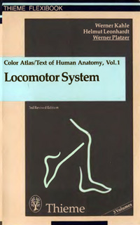
Color atlas and textbook of human anatomy. Vol.1: locomotor system
| Автор(ы): | Kahle W., Leonhardt H., Platzer W.
06.10.2007
|
| Год изд.: | 1986 |
| Описание: | A well-balanced combination of a full-fledged anatomic atlas and textbook, eminently useful to students and medical practitioners alike. Skilful visual approach to anatomy, which is a must in every physician's education, is happily wedded to a lucid text juxtaposed page by page to magnificent multicolor illustrations in such a manner that the concise description of the functional aspects of anatomy provideslet a useful guide for the perceptive student. Aspects of physiology and biochemistry are included to the extent they have a bearing on the material presented. |
| Оглавление: |
 Обложка книги.
Partsof the Body [2] General Terms [2] Principal Axes [2] Principal Planes [2] Directions in Space [2] Directions of Movement [2] Cells [4] Cytoplasm [4] Cell Nucleus [6] Vital Function of Cells [6] Tissues [8] Epithelial Tissue [8] Connective and Supporting Tissues [10] Connective Tissue [10] Cartilage [12] Bone [14] Development of Bone [16] Muscle [18] General Features of the Skeleton [20] Joints Between Bones [22] Continuous Joints Between Bones [22] Discontinuous Joints Between Bones [24] General Myology [30] Auxiliary Features of Muscles [32] Investigation of Muscle Function [32] Systematic Anatomy of the Locomotor Apparatus [35] Vertebral Column [36] Cervical Vertebrae [36] Thoracic Vertebrae [40] Lumbar Vertebrae [42] Sacrum [46] Coccyx [48] Ossification of Vertebrae [52] Intervertebral Disks [54] Ligaments of the Vertebral Column [56] Joints of the Vertebral Column [58] Vertebral Column Considered as a Whole [62] Thorax [64] Ribs [64] Sternum [66] Joints of the Ribs [68] Movements of the Thorax [70] Intrinsic Muscles of the Back [72] Lateral Tract [72] Medial Tract [74] Short Muscles of the Nape of the Neck [76] Body Wall [78] Thoracolumbar Fascia [78] Migrant Ventrolateral Muscles [78] Prevertebral Muscles [80] Scalene Muscles [80] Muscles of the Thorax [82] Intercostals [82] Abdominal Wall [84] Superficial Abdominal Muscles [84] Function of the Superficial Abdominal Musculature [90] Fasciae of Abdominal Wall [92] Deep Abdominal Muscles [94] Sites of Weakness in the Abdominal Wall [96] Abdominal Wall from Inside [98] Diaphragm [102] Position and Function of the Diaphragm [104] Sites of Diaphragmatic Hernias [104] Pelvic Floor [106] Pelvic Diaphragm [106] Urogenital Diaphragm [106] Bones, Ligaments, Joints [108] Scapula [108] Clavicle [110] Joints of the Shoulder Girdle [110] Humerus [112] Shoulder Joint [114] Radius [116] Ulna [116] Elbow Joint [118] Distal Radioulnar Joint [120] Continuous Fibrous Joint Between Radius and Ulna [120] Carpus [122] Individual Bones of the Carpus [124] Bones of the Metacarpus and Digits [126] Radiocarpal and Midcarpal Joints [128] Movements in the Radiocarpal and Midcarpal Joints [130] Carpometacarpal Joint of the Thumb [132] Carpometacarpal Joints [132] Intermetacarpal Joints [132] Metacarpophalangeal and Digital Joints [132] Muscles of the Shoulder Girdle and Upper Arm [134] Classification of the Muscles [134] Shoulder Muscles Inserted on the Humerus [136] Dorsal Muscle Group [136] Ventral Muscle Group [140] Immigrant Trunk Muscles Inserting on the Shoulder Girdle [142] Dorsal Muscle Group [142] Ventral Muscle Group [144] Muscles of the Shoulder Girdle [146] Classification According to Function [146] Fascias and Spaces in the Shoulder Girdle Region [150] Fascias [150] Special Spaces in the Shoulder Region (Axillary Spaces and Axilla) [150] Upper Arm Muscles [152] Ventral Muscle Group [152] Dorsal Muscle Group [154] Forearm Muscles [156] Classification of the Muscles [156] Superficial Layer of the Ventral Forearm Muscles [158] Deep Layer of the Ventral Forearm Muscles [160] Radial Group Includes three Muscles which Act as Flexors at the Elbow Joint [162] Superficial (Ulnar) Layer of the Dorsal Forearm Muscles [164] Deep Layer of Dorsal Forearm Muscles [166] Muscles of the Elbow Joint and the Forearm [168] Classification According to Function [168] Muscles of the Hand [170] Classification According to Function [170] Intrinsic Muscles of the Hand [172] Muscles of the Metacarpus [172] Thenar Muscles [174] Palmar Aponeurosis and Hypothenar Muscles [176] Fascias and Special Features [178] Fascias [178] Tendon sheats [180] Bones. Ligaments. Joints [182] Hip Bone [182] Junctions Between the Bones of the Pelvis [184] Morphology of the Bony Pelvis [184] Orientation of the Pelvis and Sex Differences [186] Femur [188] Patella [190] Femur [192] Hip Joint [194] Tibia [198] Fibula [200] Knee Joint [202] Attidude of Lower Limb [210] Connections Between the Tibia and the Fibula [210] Bones of the Foot [212] Bones at the Foot [216] Joints of the Foot [218] Morphology and Function of the Skeleton of the Foot [222] Plantar Arch [224] Foot Shapes [226] Muscles of the Hip and Thigh [228] Classification of the Muscles [228] Hip Muscles [230] Dorsal Hip Muscles [232] Ventral Hip Muscles [234] Thigh Muscles [236] Adductors of the Thigh [238] Hip Muscles [240] Classification According to Function [240] Thigh Muscles [244] Anterior Thigh Muscles [244] Posterior Thigh Muscles [246] Muscles of the Knee Joint [248] Classification According to Function [248] Fascias of the Hip and Thigh [250] Long Muscles of the Leg and the Foot [252] Classification of the Muscles [252] Leg Muscles [254] Extensor Group [254] Peroneal Group [256] Posterior Leg Muscles. Superficial Layer [258] Posterior Leg Muscles. Deep Layer [260] Talocrural. Subtalar and Talocalcaneonavicular Joint Muscles [262] Classification According to Function [262] Intrinsic Muscles of the Foot [264] Muscles of the Dorsum of the Foot [264] Muscles of the Sole of the Foot [266] Fascias of the Leg [272] Tendon Sheaths in the Foot [274] Skull [276] Ossification otthe Skull [276] Special Features of Intramembranous Ossification [278] Calvaria [280] Lateral View of the Skull [282] Posterior View of the Skull [284] Anterior View of the Skull [286] Inferior View of the Skull [288] Interior View of the Base of the Skull [290] Common Variants of the Interior Surface of the Base of the Skull [292] Sites of Transmission for Vessels and Nerves [294] Mandible [296] Shape of Mandible [298] Hyoid Bone [298] Orbital Cavity [300] Nasal Cavity [302] Skull Shapes [304] Special Skull Shapes and Sutures [306] Accessory Bones of the Skull [308] Temporomandibular Joint [310] Muscles of the Head [312] Mimetic Muscles of the Scalp [312] Mimetic Muscles in the Region of the Palpebral Fissure [314] Mimetic Muscles in the Nasal Region [314] Mimetic Muscles in the Region of the mouth [316] Muscles of Mastication [318] Anterior Muscles of the Neck [320] Infrahyoid Muscles [320] Musclesof the Head [322] Attachment to the Shoulder Girdle [322] Fascias of the Neck [324] Topography of Peripheral Pathways [327] Head and Neck [328] Regions [328] Anterior Facial Regions [330] Orbital Region [332] Lateral Facial Regions [334] Infratemporal Fossa [336] Superior View of the Orbit [338] Occipital and Nuchal Regions [340] Suboccipital Triangle [340] Parapharyngeal and Retropharyngeal Spaces [342] Submandibular Triangle [344] Retromandibular Fossa [346] Middle Regionof the Neck [348] Thyroid Region [350] Ventrolateral Regions of the Neck [352] Scalenovertebral Triangle [360] Upper Limb [362] Regions [362] Deltopectoral Triangle [364] Axillary Region [366] Axillary Spaces [368] Anterior Brachial Region [370] Cubital Fossa [374] Anterior Antebrachial Region [378] Wrist. Palmar Surface [380] Palm of the Hand [380] Dorsum of the Hand [384] Radial foveola [384] Lower Limb [386] Regions [386] Subinguinal Region [388] Saphenous Hiatus [390] Gluteal Region [392] Anterior Femoral Region [396] Posterior Femoral Region [400] Popliteal Fossa [402] Anterior Region of the Leg [406] Posterior Region of the Leg [408] Medial retromalleolar Region [410] Dorsum of the Foot [412] Soleof the Foot [414] Literature [418] Index [426] |
| Формат: | djvu |
| Размер: | 8964613 байт |
| Язык: | ENG |
| Рейтинг: |
126
|
| Открыть: | Ссылка (RU) |